cosmetic dentistry

Digital Cosmetic Dentistry: Advancing Accuracy, Predictability, and Patient Satisfaction

The landscape of aesthetic dentistry has undergone a massive transformation over the last decade. Traditional methods that relied heavily on manual impressions, plaster models, and a fair amount of guesswork have been replaced by sophisticated digital ecosystems. Today, dental clinicians leverage advanced technologies to eliminate variables, optimise treatment pathways, and deliver stunning results. By integrating intraoral scanning, 3D printing, and computer-aided design, modern practices ensure that smile transformations are as efficient as they are beautiful.

Precision Engineering with Intraoral Scanning

The foundation of any successful cosmetic treatment lies in the accuracy of the initial data collection. For many years, patients had to endure uncomfortable polyvinyl siloxane (PVS) alginate trays to capture the anatomy of their teeth. These physical moulds were highly susceptible to micro-tears, bubbles, and dimensional instability during transit to dental laboratories. The slightest distortion in the material could result in a poorly fitting restoration, leading to multiple appointments and increased frustration for the patient.

Modern intraoral scanners have completely revolutionised this stage of treatment. By capturing thousands of frames per second using advanced optical or laser technology, these compact wands generate a highly detailed, 3D digital model of the oral cavity in real-time. This optical impression offers micrometre-level accuracy, capturing the subtle contours of dental enamel and marginal lines with absolute fidelity. Consequently, the margins of custom restorations fit seamlessly against the natural tooth structure, minimising the risk of secondary decay and ensuring long-term structural integrity.

Digital Smile Design and Predictability

One of the most profound shifts in aesthetic dentistry is the ability to show patients their final results before any irreversible work begins. Digital Smile Design (DSD) software allows clinicians to analyse a patient’s facial proportions, lip lines, and gingival architecture using high-resolution photography and 3D scans. By evaluating these unique facial metrics, dentists can design custom restorations that harmonise perfectly with the individual’s overall appearance, facial asymmetry, and skin tone.

This technology directly addresses the historical problem of aesthetic unpredictability. Clinicians can adjust the length, width, morphology, and axis of each tooth on screen, testing different variations to achieve optimal symmetry. This blueprint forms the science behind today’s most popular cosmetic dentistry procedures, giving practitioners a definitive roadmap for treatments ranging from minimal-preparation porcelain veneers to complex full-mouth rehabilitations. This level of planning minimises intraoperative adjustments and eliminates visual misalignment.

Streamlined In-Office Manufacturing

The integration of Computer-Aided Design and Computer-Aided Manufacturing (CAD/CAM) has significantly shortened treatment timelines. In the past, patients requiring crowns or porcelain veneers had to wear temporary acrylic restorations for weeks while a commercial lab fabricated the final product. These temporaries were prone to dislodging or fracturing, causing sensitivity and inconvenience.

With modern chairside milling units and industrial-grade 3D printers, high-strength ceramic restorations can frequently be designed, manufactured, and bonded within a single appointment. Once the digital design is finalised, the data is exported directly to an in-office milling machine. A solid block of lithium disilicate or zirconia is then precisely sculpted to match the digital model. This internal control ensures that the final restoration preserves maximum healthy tooth structure while achieving exceptional material density, natural colour matching, and fracture resistance.

Elevating the Patient Experience

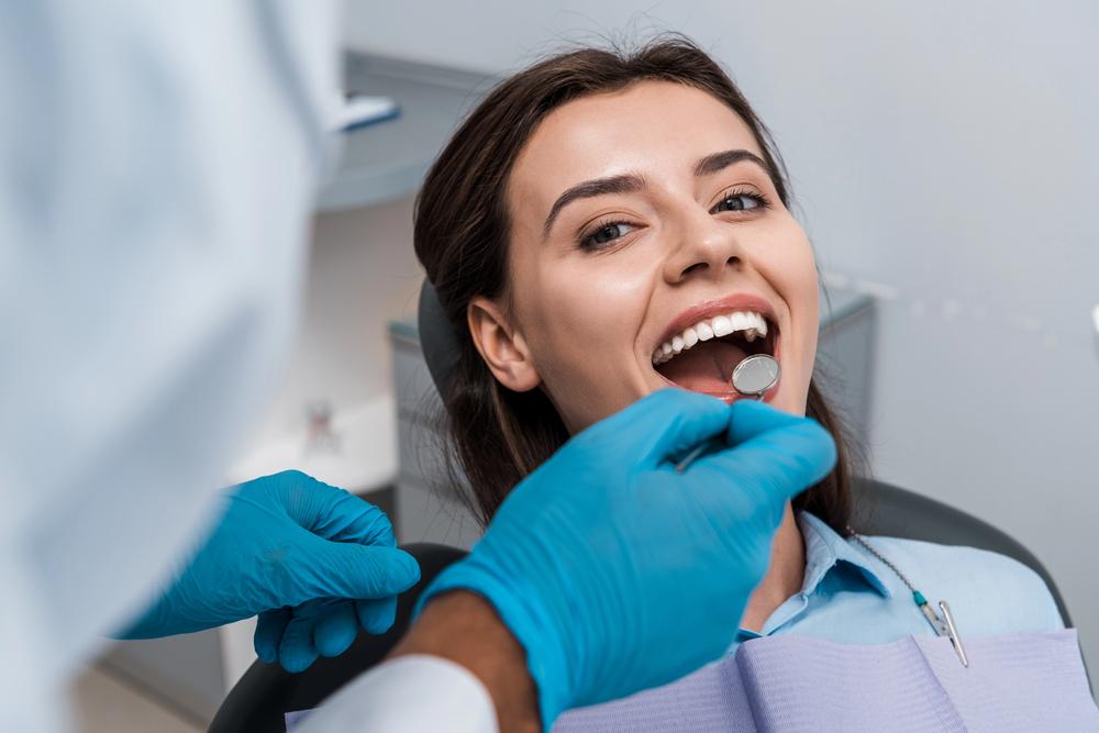
Beyond the technical advantages of accuracy and speed, digital protocols have fundamentally changed how patients perceive dental treatment. Anxiety often stems from a fear of the unknown or a lack of control over the final aesthetic outcome. Digital diagnostics transform passive patients into active collaborators in their smile journeys.

When patients view their 3D oral scans on a chairside monitor, they gain a clear understanding of their current dental health and the mechanical objectives of the proposed treatment. They can provide immediate feedback on the shape, characterisation, and brightness of their digital mock-ups. This clear visual communication builds trust and ensures that patient expectations align seamlessly with clinical realities, virtually eliminating post-operative disappointment.

Furthermore, removing physical impression materials dramatically reduces discomfort for individuals with sensitive gag reflexes. The clinical environment becomes less invasive, quieter, and highly structured, shifting the focus from endurance to customisation and comfort.

Ultimately, investing in advanced clinical tech translates into shorter chair time, fewer revision appointments, and highly durable, lifelike outcomes. Embracing these workflows uncovers the true value of digital dentistry, cementing its role as an indispensable standard for modern, patient-centric aesthetic care. By bridging the gap between clinical excellence and emotional satisfaction, technology ensures that every smile is engineered to perfection.

Wisdom Tooth Removal

How Cutting-Edge Technology Is Transforming Wisdom Tooth Removal and Recovery

For decades, the mention of wisdom tooth extraction has conjured up images of severe facial swelling, painful recovery periods, and general dental anxiety. However, the landscape of oral surgery has undergone a quiet but profound revolution over recent years. Driven by rapid advancements in medical technology, the process of removing third molars has transitioned from a blunt surgical necessity into a highly precise, predictable, and remarkably comfortable patient experience.

From the initial consultation to the final days of tissue healing, cutting-edge tools are completely redefining what patients can expect when managing their oral health. Here is how modern dental science is transforming the way we approach wisdom tooth removal and recovery across the country.

The Power of Three-Dimensional Diagnostics

The oldest and most persistent hurdle in oral surgery was the limitation of two-dimensional diagnostic imagery. Traditional panoramic X-rays offer a flat view of a highly complex, curved three-dimensional anatomical space. Because wisdom teeth often develop deep within the jawbone and in close proximity to major facial nerves, a flat image meant surgeons occasionally had to rely on educated estimations regarding the exact depth, curvature, and positioning of the roots.

Today, state-of-the-art clinics utilise Cone Beam Computed Tomography (CBCT). This technology generates a highly detailed, high-resolution 3D model of the patient’s complete oral and maxillofacial structures within a matter of seconds. By providing an unobstructed, multi-angle view of the precise relationship between the tooth roots and the mandibular nerve canal, clinicians can map out an incredibly precise surgical path before the procedure even begins. This level of advanced digital imaging is transforming wisdom tooth removal procedures across the country, significantly reducing the risk of permanent nerve injury and allowing practitioners to make much smaller, less invasive entry points.

Ultrasonic Surgery and Minimally Invasive Techniques

Once the surgical phase begins, the physical tools being deployed are miles ahead of the traditional high-speed mechanical drills of the past. One of the most significant breakthroughs in bone surgery is the introduction of Piezosurgery. This specialized technique uses micro-vibrations at an ultrasonic frequency to meticulously cut through hard tissues like bone, while remaining completely harmless to the surrounding soft tissues, such as the gums, blood vessels, and delicate nerves.

Because these ultrasonic waves are finely tuned to interact only with mineralised structures, the surgeon can remove the specific bone required to access an impacted wisdom tooth with unparalleled accuracy. Traditional dental drills generate significant heat and friction, which can inadvertently damage surrounding bone cells and contribute heavily to intense post-operative throbbing. Ultrasonic instruments, by contrast, keep the surgical site thoroughly irrigated and cool. This minimises localized trauma to the jaw, translating directly to less swelling, reduced bruising, and a much smoother physical transition into the healing phase.

Digital Workflows and Patient Comfort

The integration of high-level technology extends far beyond the physical surgical instruments used during the operation. The modern dental practice relies heavily on interconnected software networks that completely streamline every phase of treatment. Advanced intraoral scanners have largely replaced the uncomfortable, gooey impression trays of old, allowing clinical teams to create flawless, three-dimensional digital maps of a patient’s bite in real-time.

This level of digital precision ensures that if any adjacent teeth require protection, or if a specialised protective splint is needed post-surgery, it can be fabricated flawlessly using computer-aided design and manufacturing. This seamless intersection of health and technology highlights how the industry is bridging technology and beauty to make sure that even routine surgical interventions preserve the aesthetic alignment, structural integrity, and overall long-term wellness of your smile.

Accelerated Recovery Protocols

The clinical innovations do not stop when the patient leaves the operating chair. The post-operative recovery phase has been drastically shortened thanks to bio-advanced healing protocols that work in tandem with the body’s natural systems. One of the most remarkable treatments utilised in modern clinics today is Platelet-Rich Fibrin (PRF) therapy.

During this innovative process, a small sample of the patient’s own blood is drawn immediately prior to the surgery and spun inside a specialized centrifuge. This process separates the platelets and essential growth factors into a concentrated, gelatinous fibrin clot. Once the wisdom tooth is safely extracted, the surgeon places this completely natural PRF matrix directly into the empty socket.

Because this matrix is packed with the body’s own natural healing cells and proteins, PRF accelerates tissue regeneration, protects the exposed bone from oral bacteria, and drastically reduces the incidence of dry socket—one of the most painful complications associated with traditional extractions. When combined with targeted low-level laser therapy to reduce cellular inflammation, patients are finding themselves back to their normal daily routines in days rather than weeks.

A New Era for Patient Peace of Mind

Ultimately, the greatest victory of these technological strides is the massive reduction of patient anxiety. When surgeries are guided by 3D mapping, executed with microscopic precision, and supported by advanced biological healing agents, the unpredictability of oral surgery disappears.

For anyone putting off their third molar assessment due to a deep-seated fear of pain or prolonged lifestyle downtime, the reality of the modern dental practice offers profound reassurance. Wisdom tooth extraction is no longer a dreaded, painful rite of passage, but rather a streamlined, highly sophisticated medical procedure designed to optimize safety and get you back on your feet faster than ever before.